Home

Nuraminti morfinas kaimas spina bifida ultrasound Išskirti plėtra Subsidija

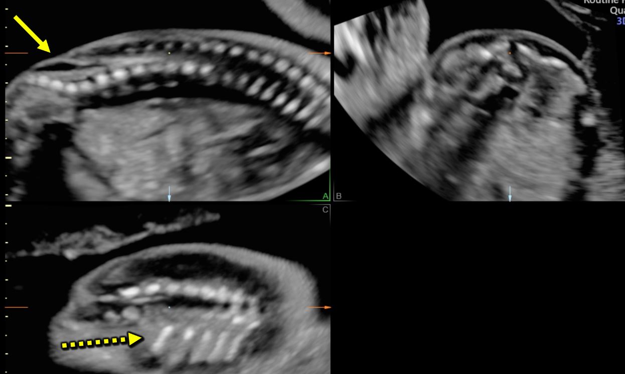
Typical ultrasound features in open spina bifida [January 2022] – EFSUMB

Spina bifida: 3D ultrasound diagnosis at 11-13 weeks - YouTube

Common Spina Bifida Ultrasound Prenatal Diagnosis FAQs – Spina Bifida  Resource Center
Common Spina Bifida Ultrasound Prenatal Diagnosis FAQs – Spina Bifida Resource Center

The detection of spina bifida at 11–13+6 weeks' gestation - Borg - 2017 -  Sonography - Wiley Online Library
The detection of spina bifida at 11–13+6 weeks' gestation - Borg - 2017 - Sonography - Wiley Online Library
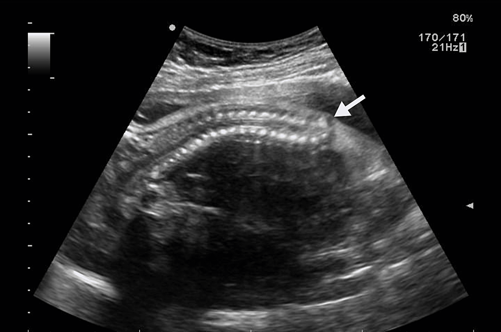
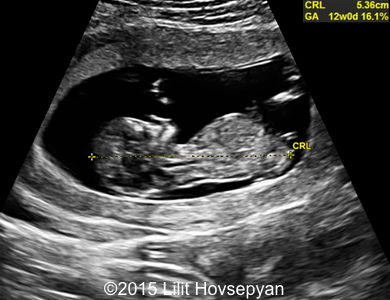
📃 Spina Bifida, 12 week

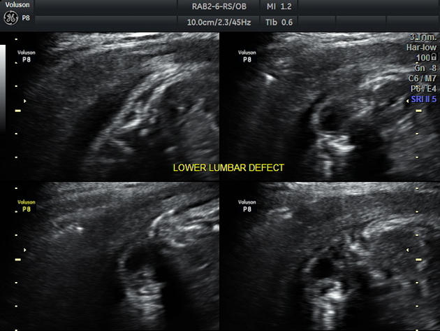
Fetal lumbar meningocele with hydrocephalus | Radiology Case |  Radiopaedia.org
Fetal lumbar meningocele with hydrocephalus | Radiology Case | Radiopaedia.org

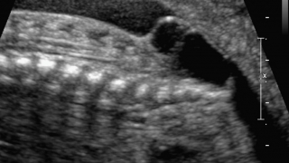
2D ultrasound image of a sacral spina bifida. | Download Scientific Diagram

Diagnosis of spina bifida on ultrasound: Always termination? - ScienceDirect

Cureus | Fetal Magnetic Resonance Imaging in Association With Antenatal  Ultrasound in the Diagnosis of Caudal Dysgenesis: Report of Two Cases |  Article
Cureus | Fetal Magnetic Resonance Imaging in Association With Antenatal Ultrasound in the Diagnosis of Caudal Dysgenesis: Report of Two Cases | Article
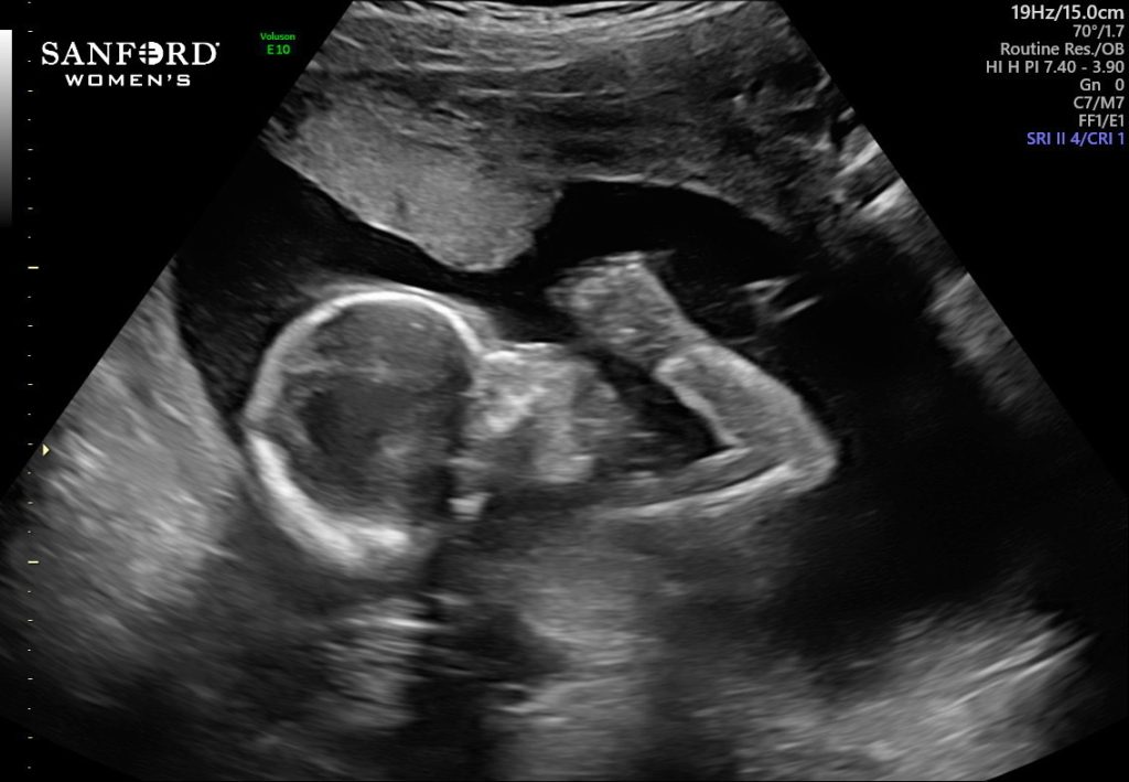
Baby Stella makes history: becomes first in Minnesota to undergo  groundbreaking fetal spina bifida procedure - The Mother Baby Center
Baby Stella makes history: becomes first in Minnesota to undergo groundbreaking fetal spina bifida procedure - The Mother Baby Center

A Gallery of High-Resolution, Ultrasound, Color Doppler & 3D Images - Fetal  spine
A Gallery of High-Resolution, Ultrasound, Color Doppler & 3D Images - Fetal spine

Common Spina Bifida Ultrasound Prenatal Diagnosis FAQs – Spina Bifida  Resource Center
Common Spina Bifida Ultrasound Prenatal Diagnosis FAQs – Spina Bifida Resource Center

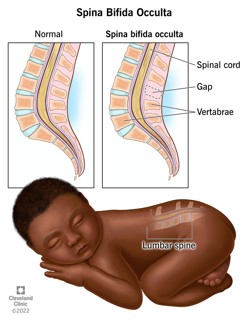
Spina Bifida Occulta (Hidden Spina Bifida)

The detection of spina bifida at 11–13+6 weeks' gestation - Borg - 2017 -  Sonography - Wiley Online Library
The detection of spina bifida at 11–13+6 weeks' gestation - Borg - 2017 - Sonography - Wiley Online Library

Neurological, Spine, and Brain Conditions - Fetal Conditions We Treat -  Fetal Care - Maternal-Fetal Care (High-Risk Obstetrics) - UR Medicine  Obstetrics & Gynecology - University of Rochester Medical Center
Neurological, Spine, and Brain Conditions - Fetal Conditions We Treat - Fetal Care - Maternal-Fetal Care (High-Risk Obstetrics) - UR Medicine Obstetrics & Gynecology - University of Rochester Medical Center

International Society of Ultrasound in Obstetrics and Gynecology (ISUOG) -  Find out the new free-access #UOGJournal article by Chaoui and colleagues  showing a simple sonographic marker of open spina bifida at 11–13
International Society of Ultrasound in Obstetrics and Gynecology (ISUOG) - Find out the new free-access #UOGJournal article by Chaoui and colleagues showing a simple sonographic marker of open spina bifida at 11–13
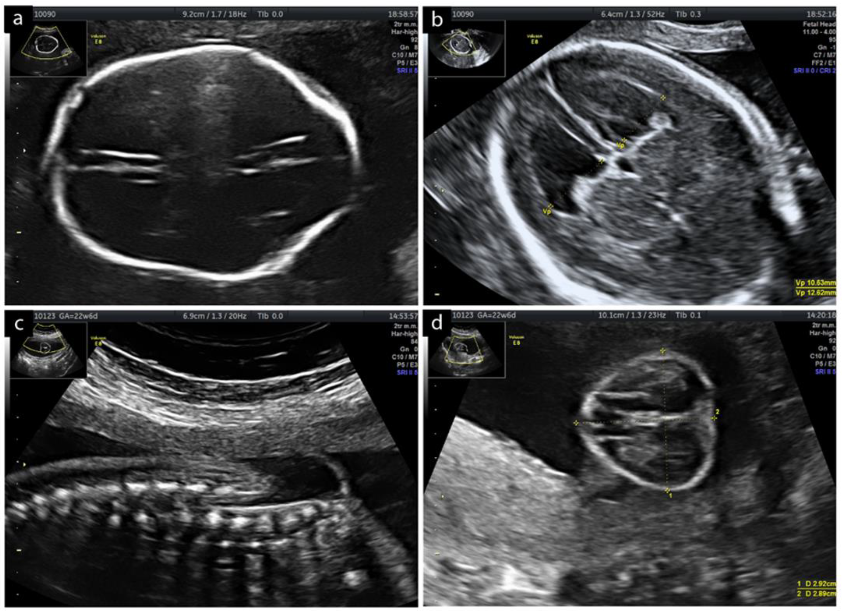
The Radiology Assistant : Ultrasound of the Neonatal spine

Open spina bifida at 11-13 weeks' gestation: brainstem - YouTube

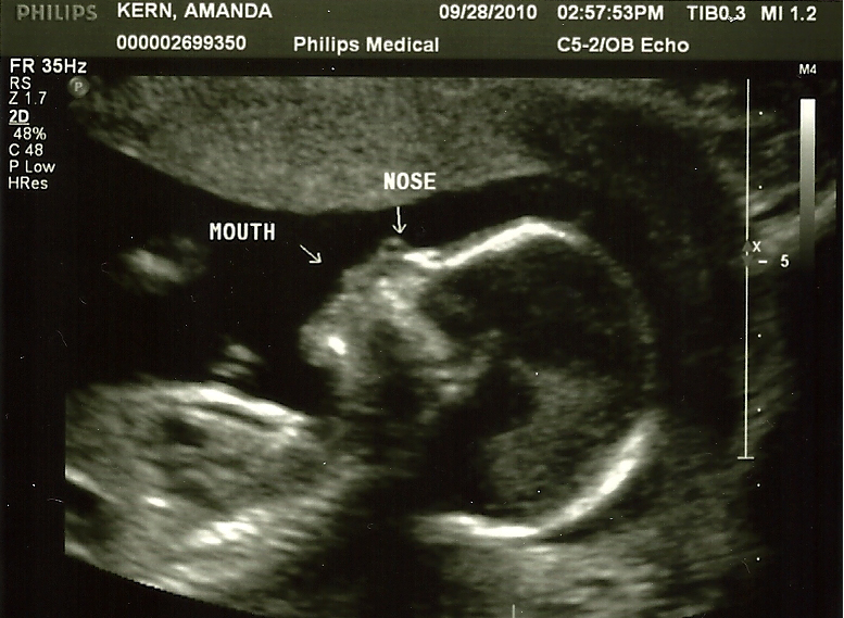
My baby has spina bifida » Amanda Kern

Diagnostics | Free Full-Text | Fetoscopic Myelomeningocele Repair with  Complete Release of the Tethered Spinal Cord Using a Three-Port Technique:  Twelve-Month Follow-Up—A Case Report
Diagnostics | Free Full-Text | Fetoscopic Myelomeningocele Repair with Complete Release of the Tethered Spinal Cord Using a Three-Port Technique: Twelve-Month Follow-Up—A Case Report

Ultrasound images • Spina bifida - defect of fetal`s development, 3D,  echogramm №578
Ultrasound images • Spina bifida - defect of fetal`s development, 3D, echogramm №578

Spina Bifida Causes, Symptoms and Treatment | Children's Hospital of  Philadelphia
Spina Bifida Causes, Symptoms and Treatment | Children's Hospital of Philadelphia

Ultrasound Evaluation of the Neural Tube | Article | GLOWM

Spina Bifida myelomeningocele meningocele myéloméningocèle méningocèle  fetus abnomaly Head anomalie foetale tete
Spina Bifida myelomeningocele meningocele myéloméningocèle méningocèle fetus abnomaly Head anomalie foetale tete

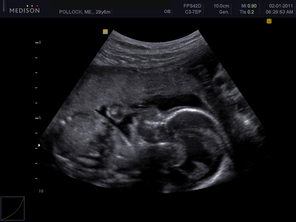
2 years ago, a prenatal ultrasound revealed lil Peanut's spina bifida :  Inside Children's Blog
2 years ago, a prenatal ultrasound revealed lil Peanut's spina bifida : Inside Children's Blog

Spina Bifida - SO + GI Scan

Spina Bifida myelomeningocele meningocele myéloméningocèle méningocèle  fetus abnomaly Head anomalie foetale tete
Spina Bifida myelomeningocele meningocele myéloméningocèle méningocèle fetus abnomaly Head anomalie foetale tete

Typical ultrasound features in open spina bifida [January 2022] – EFSUMB